 Diagnostics
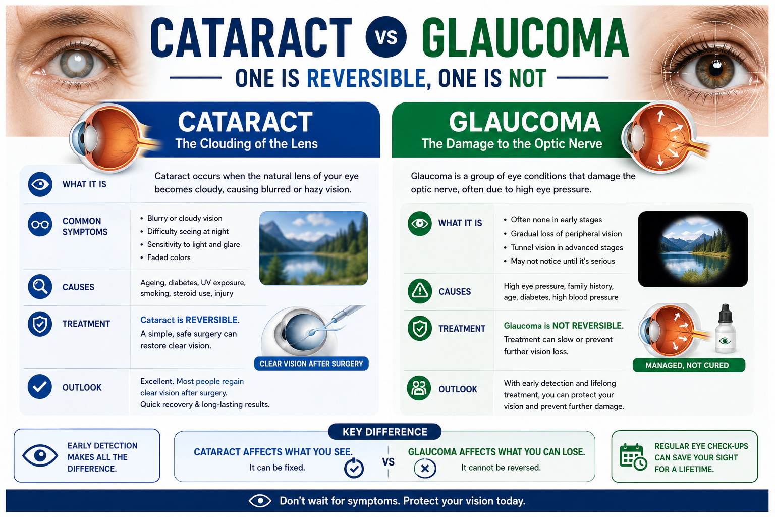
OCT (Optical Coherence Tomography)
What it examines: Cross-sectional imaging of the retina and optic nerve layers.
Why a specialist may recommend it: Detects macular swelling, glaucoma-related nerve thinning and retinal disease before symptoms appear.
What you can expect: Non-contact light scan; a few minutes per eye. Dilatation may be advised.
 Diagnostics
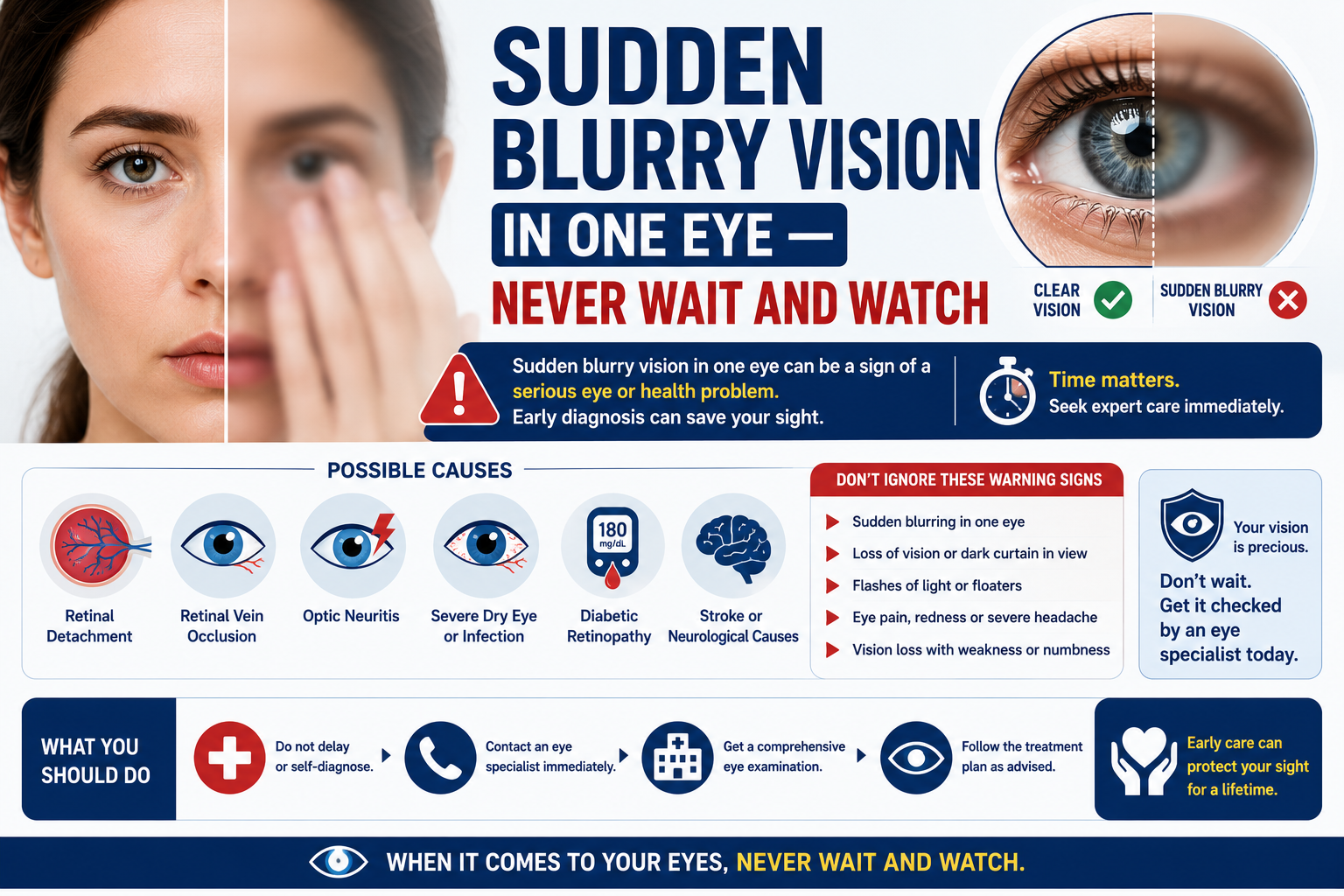
Corneal Topography
What it examines: A curvature and elevation map of the cornea.
Why a specialist may recommend it: Essential for LASIK suitability, keratoconus detection and contact-lens fitting.
What you can expect: Painless; you simply look at a target light for a few seconds.
 Diagnostics
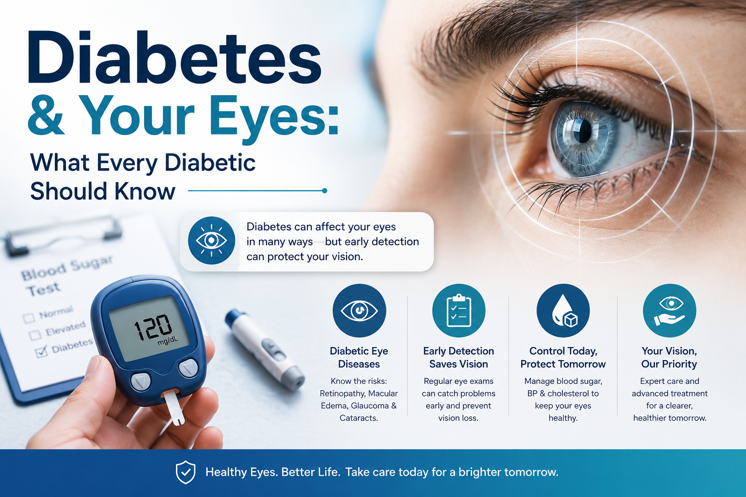
Fundus Imaging
What it examines: High-resolution photographs of the retina.
Why a specialist may recommend it: Documents diabetic changes, retinal tears and other findings for accurate comparison over time.
What you can expect: A bright flash photograph; usually quick, sometimes after dilatation.
 Diagnostics
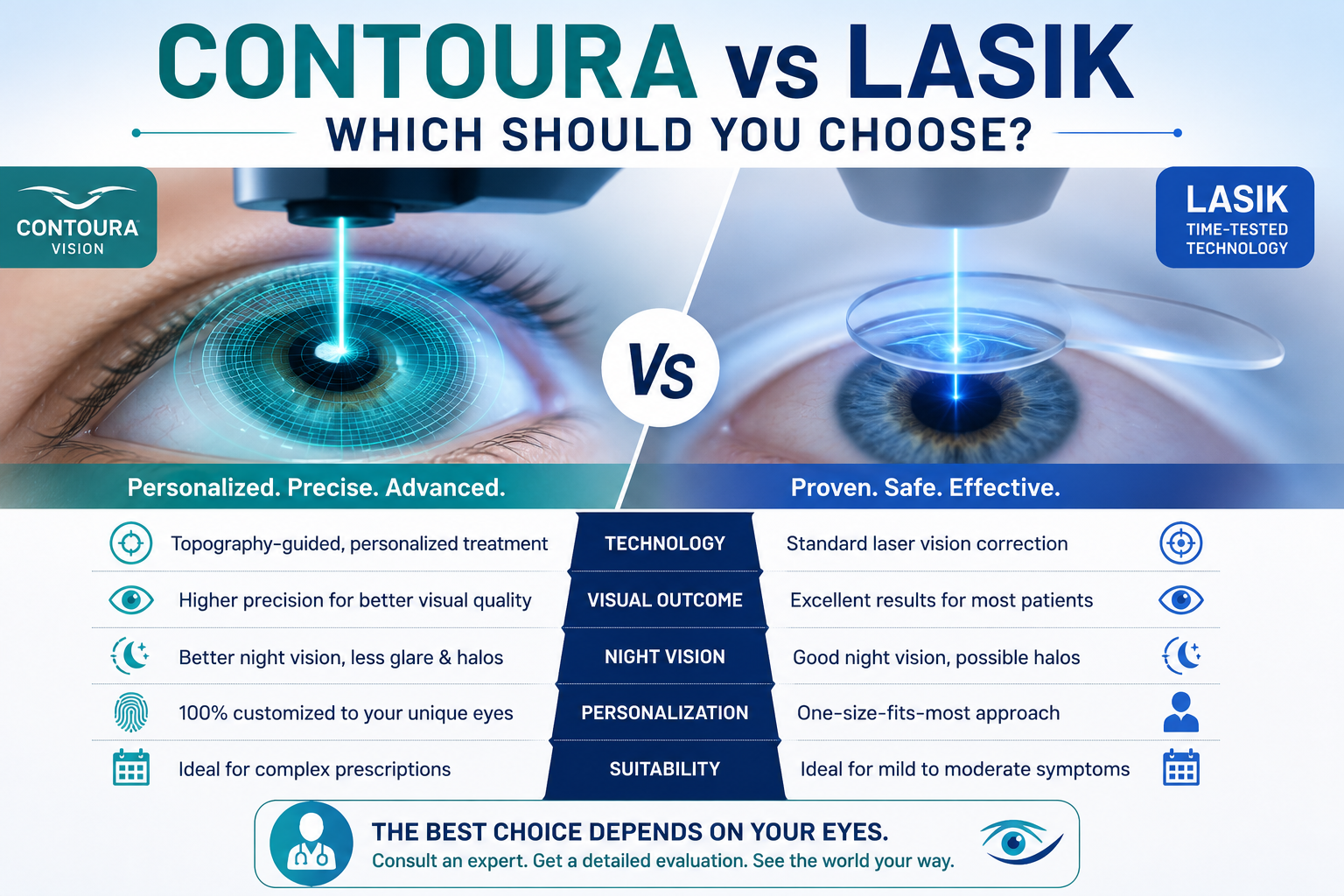
Computerised Perimetry
What it examines: Maps your field of vision point by point.
Why a specialist may recommend it: Detects and monitors glaucoma and neurological visual field loss.
What you can expect: You press a button when you see lights; typically 10–15 minutes.
 Diagnostics
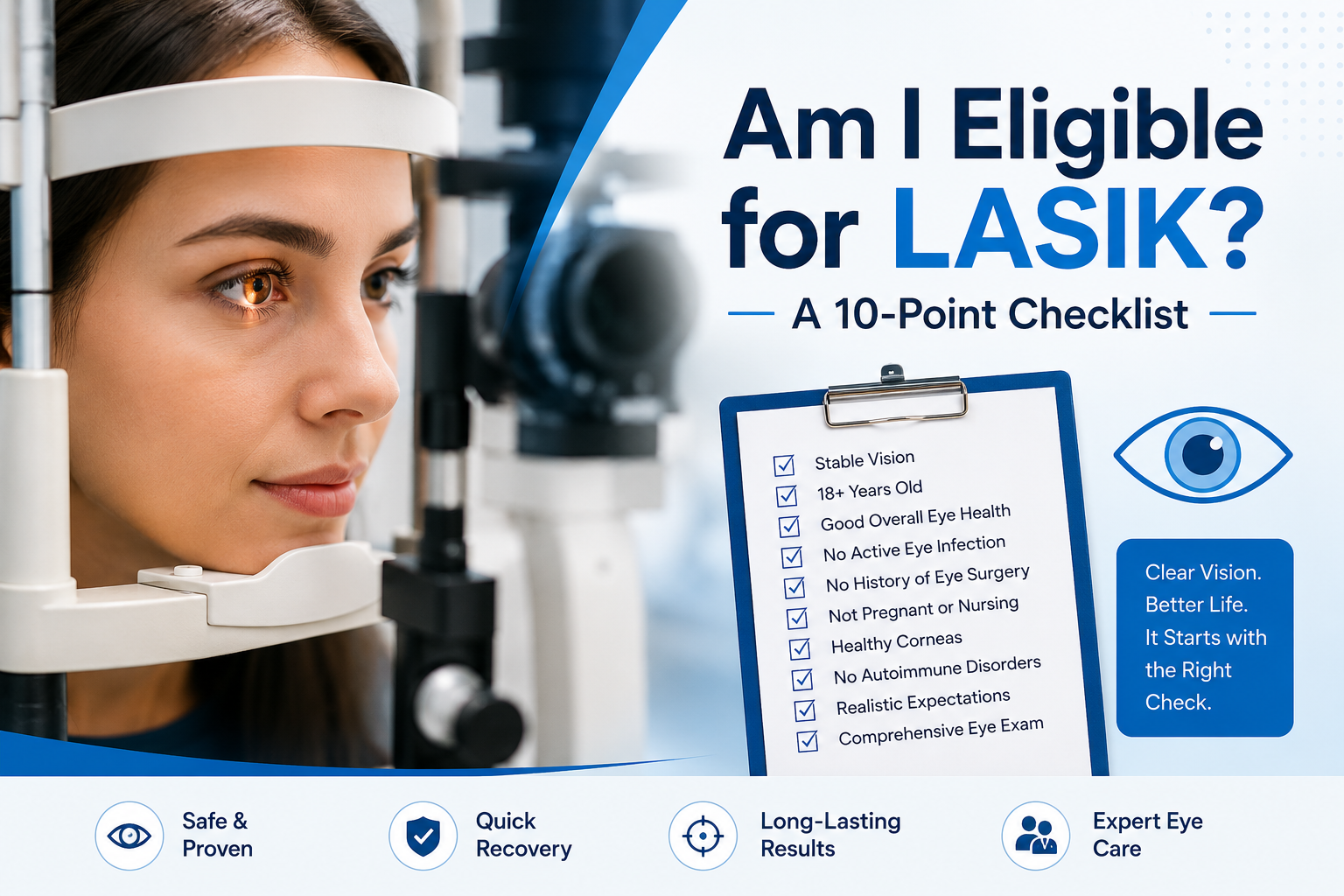
Slit-lamp Examination
What it examines: Microscope examination of the front and back of the eye.
Why a specialist may recommend it: The foundation of every eye examination — detects cataract, corneal and surface disease.
What you can expect: You rest your chin on the microscope; completely non-invasive.
 Diagnostics
Eye Pressure Assessment (Tonometry)
What it examines: Measures intraocular pressure.
Why a specialist may recommend it: A key screening and monitoring test for glaucoma.
What you can expect: A gentle puff of air or a numbed touch measurement; seconds per eye.
Equipment availability and test durations are confirmed at the time of booking. Visual guides are for patient education.